

Ultrasound Biomicroscopy
What is Ultrasound Biomicroscopy?
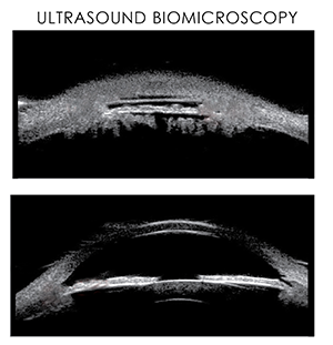
Ultrasound biomicroscopy is an eye examination that provides a detailed image of your eye. It uses high-energy sound waves to generate images of the inside of the eye. Ultrasound biomicroscopy is most commonly used to detect glaucoma (increase in eye pressure and damage of optic nerves), cysts, tumours, foreign objects in the eye and extent of trauma.
During the examination, you will be asked to lie down in a supine (face-up) position. Your examiner will apply topical anaesthesia between the eyelids before scanning. The probe of the ultrasound is placed on the eyeball. Ultrasound biomicroscopy generates cross-sectional or transverse images of the following structures of the eye:
- Cornea: Transparent, dome-shaped layer covering the front of the eye
- Sclera: The white portion of the eye
- Iris: Thin, circular structure that regulates the amount of light that enters the eye
- Anterior chamber angle: The angle formed between the iris and the cornea
- Ciliary body: Releases transparent fluid called aqueous humour, which maintains normal eye pressure
The images help your doctor to understand the structural pattern of the eye and diagnose any abnormalities or problems.