

Retinal Tear
What is Retinal Tear?
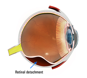
The retina is tissue present at the back of the eye that is responsible for vision. Severe near-sightedness, eye injury, cataract surgery or familial history may cause a retinal tear. The retina is attached to the choroid tissue which provides the blood supply to the retina. When the retina tears, it gets detached from the choroid tissue along with its blood supply. A retinal tear is characterized by symptoms such as the appearance of floaters (small moving spots in the vision field), flashes of light in front of the eyes, darkening of your side vision and gradual loss of vision.
Detachment of the retina and retinal tears can be diagnosed by a complete and thorough check-up of your eye. Your doctor may gently apply pressure on your eye with a blunt instrument or cotton swab, and shine a bright light to examine the retina. Additional eye tests may be ordered, such as eye pressure test, vision test, and ultrasound, to determine the cause of symptoms.
Treatment for Retinal Tears
Retinal tears can be treated if diagnosed in the early stages. The various methods of treatment include:
- Laser surgery: Laser beams are used to create burns. The scars formed help to seal the tear.
- Freeze treatment: A freezing probe is used to freeze the tissue around the tear. The scars formed help to seal the tear.
- Pneumatic retinopexy: In this procedure, a gas bubble is injected into the eye. It pushes against the retina and closes the tear. The tear is then sealed by freezing or using a laser.
- Scleral buckle: a silicone band is stitched onto the white of the eye so that it pushes against the tear till it heals.
- Vitrectomy: This procedure is used for large retinal tears. Your doctor replaces your vitreous (a gel-like substance between the lens and the retina) with a saline solution.
Related Topics:
- Uveitis and Ocular Inflammation
- Dry Eyes
- Lid Cysts
- Blepharitis
- Glaucoma
- Retinal Tear
- Cataract
- Diabetic Macular Oedema
- Retinal Vein Occlusion
- Macular Oedema
- Cystoid Macular Oedema
- Central Serous Retinopathy
- Vision Disorders
- Watery Eye
- Tear Duct Obstruction
- Vein Occlusion
- Chalazion
- Vein Occlusion Macular Oedema
- Allergic Disorders of the Eye
- Blurred Vision
- Distortion of Central Vision
- Ocular Ischemic Syndrome
- Optic Neuropathy
- Posterior Uveitis
- Proliferative Diabetic Retinopathy
- Temporal Arteritis
- WET AMD
- Traumatic Iritis
- Acute/ Chronic/Recurrent Iridocyclitis
- Am I at Risk of Glaucoma?
- Epiretinal Membrane
- Open and Closed Iridocorneal Angles
- Pars Planitis/Intermediate Uveitis
- Retinal Detachment
- Subconjunctival Haemorrhage