

Vein Occlusion
What is Vein Occlusion?
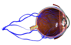
The retina is the layer of tissue present at the back of the eye. It changes the light that enters the eye into nerve signals, which are transmitted to the brain for interpretation. Blood vessels supply oxygen and nutrients to the retina to keep it in working condition. As you age, atherosclerosis (hardening or thickening) and clot formation can occur in these vessels. These blocked arteries cross over retinal veins, exerting pressure on them and disrupting the flow of blood. A block in these veins that carry blood away from the retina can lead to a condition known as retinal vein occlusion (RVO).
There are two types:
- Central retinal vein occlusion (CRVO): The block occurs in the central/main retinal vein.
- Branch retinal vein occlusion (BRVO): The block occurs on the surface of the retina, where the main retinal vein branches into smaller veins.
Older people are most often affected by retinal vein occlusion. The condition may be painless and often occurs in only one eye.
Symptoms of Vein Occlusion
Sudden blurring or loss of vision, which worsens over time. Some patients may experience the complete loss of vision
Risk factors for Vein Occlusion
Some of the risk factors for retinal vein occlusion include:
- Smoking
- Hypertension
- Diabetes
- Hypercholesterolemia
- Atherosclerosis
- Other eye conditions such as glaucoma.
Treatments for Vein occlusion
Vein occlusion can be treated with:
- Lucentis Injection
- Ozurdex
- Macular Laser
- Panretinal Photocoagulation
When left untreated, retinal vein occlusion can lead to further complications such as macular oedema (fluid buildup in the macula, present in the centre of the retina) and neovascularization (growth of new abnormal blood vessels in the retina) leading to further worsening of vision.
Related Topics:
- Uveitis and Ocular Inflammation
- Dry Eyes
- Lid Cysts
- Blepharitis
- Glaucoma
- Retinal Tear
- Cataract
- Diabetic Macular Oedema
- Retinal Vein Occlusion
- Macular Oedema
- Cystoid Macular Oedema
- Central Serous Retinopathy
- Vision Disorders
- Watery Eye
- Tear Duct Obstruction
- Vein Occlusion
- Chalazion
- Vein Occlusion Macular Oedema
- Allergic Disorders of the Eye
- Blurred Vision
- Distortion of Central Vision
- Ocular Ischemic Syndrome
- Optic Neuropathy
- Posterior Uveitis
- Proliferative Diabetic Retinopathy
- Temporal Arteritis
- WET AMD
- Traumatic Iritis
- Acute/ Chronic/Recurrent Iridocyclitis
- Am I at Risk of Glaucoma?
- Epiretinal Membrane
- Open and Closed Iridocorneal Angles
- Pars Planitis/Intermediate Uveitis
- Retinal Detachment
- Subconjunctival Haemorrhage